Internal medicine
We also offer diagnostics and appropriate therapy for various clinical pictures in the field of internal medicine. These include, for example:
Gastroscopy (= endoscopic reflection of the esophagus and stomach)
Gastroscopy can be used to diagnose inflammatory, degenerative and anatomical changes of the esophagus and stomach. Indications for gastroscopy include stomach discomfort, previously identified stomach ulcers, recurrent colic, and pharyngeal obstruction.
Lung examination
In addition to the detailed clinical examination of the respiratory tract, we have other diagnostic tools at our disposal, such as blood gas analysis. Here, the concentration of blood gases in the horse’s body at rest and under stress can be determined directly at our clinic.
To obtain an all-encompassing view of the lungs, appropriate radiographs should also be obtained.
Sampling (biopsy) of lung tissue is also possible.
Bronchoscopy
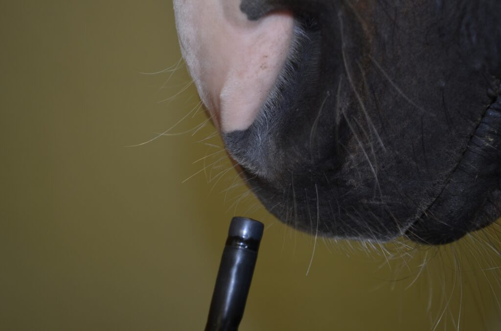
During a bronchoscopy, a tube with a camera is advanced through the nostril into the following structures. The following areas can be imaged during this procedure:
- Nasal passages: polyps or tumors may be present here, for example.
- Ethmoid bone: e.g. to detect ethmoid bone hematomas
- Air sacs: e.g. in case of air sac suppuration or fungi in the air sac
- Larynx: for clarification of functionality
- Palate: e.g. in case of cleft palate or displacement of the soft palate
- Trachea: e.g. to determine if the horse has mucus in the trachea and to take a sample if necessary.
- Branching into the main bronchi: swelling may be present here
During bronchoscopy, mucus (tracheobronchial secretion, TBS for short) is usually taken from the trachea and the cells contained therein are examined. If there is no mucus in the trachea, an irrigation sample can be obtained from the bronchi (bronchoalveolar lavage, or BAL).
Heart examinations
Electrocardiogram (ECG)
With the help of an electrocardiogram (ECG), all excitation formations as well as rhythm disturbances of the atria and the ventricles can be displayed. We use the ECG in performance diagnostics, in the individual care of our cardiological patients and in daily anesthesia monitoring.
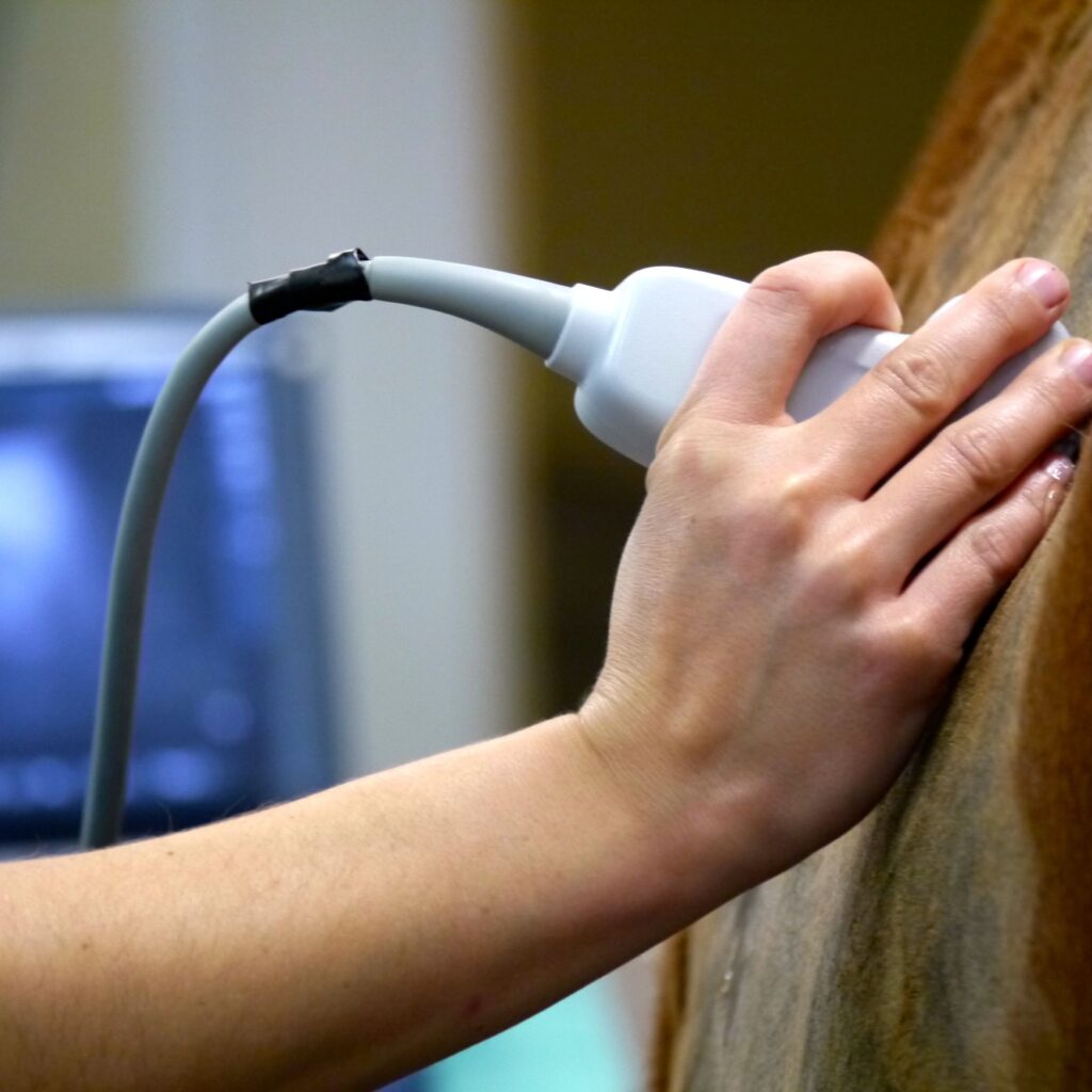
Echocardiography (“heart ultrasound”)
The examination of the heart by ultrasound is called echocardiography. Here, not only the “normal” ultrasound is used, but also the so-called Doppler. The latter makes it possible to visualize the blood flow.
Echocardiography allows the size of the individual heart chambers, the atria and the pumping function of the heart to be assessed. The function of the individual heart valves can also be shown by means of the heart ultrasound.